Is Future tech Driving Dermatology's Evolution?
An Overview of the Dermatology Market
Dermatology, like other medical fields, has witnessed and continues to experience massive technological advancements, medical advances, and procedures. Dermatologists and other skincare specialists attribute the majority of this modernization to medical advancements. Individuals can find relief from age-old skin conditions such as acne and scarring as a result of modern skincare innovations. There is a rising tide of interest in anti-aging treatments for skin.
Dermatology, like many other fields, has undergone significant transformations over the past ten years due to the development of new technologies. The implementation of refined electronic medical records (EMR) software to promote telemedicine services, is one of the most significant changes.
5 Emerging Technologies in Dermatology
- Machine learning
- Dermoscopy
- Optical Coherence Tomography
- Fluorescence Imaging
- Artificial Intelligence
Machine Learning
The incorporation of machine-based learning, a subset of artificial intelligence, into a variety of emerging imaging modalities represents a significant advancement in medical science. Variable pixel intensities in a high-resolution database of known diagnoses and treatments are comprehended by a computer. This pixel variation is utilized by the computer to analyze unspecified digital files. Computer algorithms can make more precise clinical diagnoses, sometimes even more precise than dermatologists. This is possible, however, if a large image database is utilized. The algorithm can use clinical photographs, dermoscopic images, and histopathological images to make an accurate diagnosis.
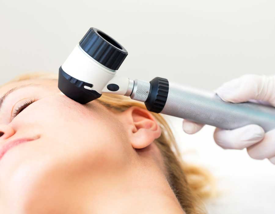
Dermoscopy
Dermoscopy, also known as an epiluminescence microscope and used as a dermatologist's stethoscope, has been shown to be an excellent diagnostic tool, not only for distinguishing melanoma from other melanocytic lesions but also for assisting in clinical diagnoses of provocative dermatoses. The fundamental principle appears to be lesion illumination and transillumination, which facilitates the visualization of fine surface and subsurface structures. The most recent generation of dermoscopic incorporates crossed polarizers, which filter out peripheral scattered light, reduce glare, and facilitate the visualization of substratal structure in the absence of linkage fluid. In addition to built-in photography and software to capture, store, and analyze images, modern dermoscopy also features integrated photography. Advanced devices with whole-body mapping systems aid in detailed analysis and follow-up.
Ex-vivo dermoscopy improves histopathological correlation, patient-physician communication, dermoscopy in aesthetic dermatology and dermatosurgery for assessing outcomes of laser hair reduction, consequences of hair transplantation, as well as for early diagnosis of post-transplant abnormalities such as folliculitis. Compared to melanoma diagnosis, it has a high sensitivity but low specificity, even when performed by experts. It is difficult to perform in some areas, such as the mucosa and does not eliminate the need for a biopsy entirely. Additionally, it requires adequate training.
Tele-dermoscopy is the transfer of digitalized dermoscopic images for diagnosis, treatment, education, and follow-up. Its advantages included fewer unnecessary referrals, shorter wait times, and cheaper skin care. The same type of service is provided by a smartphone and a mobile dermoscopy. This not only improves the accuracy of telediagnosis but also the delivery of skincare services, especially in regions with limited access to advanced medical care.
Optical Coherence Tomography
The use of OCT (optical coherence tomography) in dermatological research is growing in popularity. A crucial dermatological parameter that can be inferred from an OCT image is epidermal thickness, which can serve as an indicator of skin disease. The second important parameter is the optical resolution, which represents a lower limit on the spatial details of skin structures. OCT is performed in real-time and is capable of rapidly capturing images (1 minute). In this experiment, the echo time delay and intensity of backscattered light from tissue microstructures are measured. OCT is based on the interferometry principle developed by Michelson over a century ago. Its inability to detect individual cells restricts its diagnostic capabilities. The devices are expensive and require advanced training. In the future, multiphoton tomography could be used to enhance the visibility of cellular structures.
Fluorescence Imaging
Using the fluorescent properties of endogenous/exogenous fluorophores that absorb energy from ultraviolet/visible light radiation, fluorescence imaging (FI) generates images. Multiphoton microscopy employs a near-infrared laser. This method employs fluorescence produced by the simultaneous absorption of two photons with lower energy and longer wavelength. It facilitates the examination of crucial molecular events in cells and in vivo. It can recognize dermal collagen. It has been used to treat, among other conditions, skin cancer, aging, and inflammatory dermatoses.
Autofluorescence and quenched activity-based imaging are the two most prominent FI modalities. The former is used to identify NMSC because they contain more tryptophan residues, resulting in increased fluorescence, which allows the physician to make the diagnosis.
Artificial Intelligence
Due to its extensive clinical, dermoscopic, and histopathological image database, dermatology has taken the lead in the implementation of artificial intelligence. Machine learning is a subset of artificial intelligence in which a computer learns by creating numerous algorithms to solve problems and generate an output. There is either supervision, semi-supervision, or no supervision. AI is now widely used to differentiate benign from malignant lesions, such as to differentiate benign nevi from malignant melanoma. In addition, it is utilized to predict the complexity of malignant melanoma and aid in the histopathological diagnosis of the disease.
Teledermatology is one of the fastest-growing fields, especially in regions with limited access to skin specialists and advanced medical care. Smartphone applications are incorporating AI to image lesions, analyze them, and generate necessary referrals. There are also smartphone applications for melanoma risk assessment and diagnosis. The ranges for sensitivity and specificity were 7% to 73% and 37% to 94%, respectively. With its extensive image database and AI algorithm, dermoscopy can assist dermatologists in training and identifying dermoscopic patterns, thereby facilitating more accurate clinical diagnosis.
Conclusion
As technology advances, new devices, imaging technologies, and therapeutic modalities will become more widely available. The future dermatological practice may be significantly affected by software incorporating artificial intelligence and machine learning. Newer technologies have the potential to enhance clinical acumen, reduce invasive procedures, distinguish benign from malignant lesions, and monitor treatment outcomes. These innovations may become standard diagnostic and imaging tools in the future.
